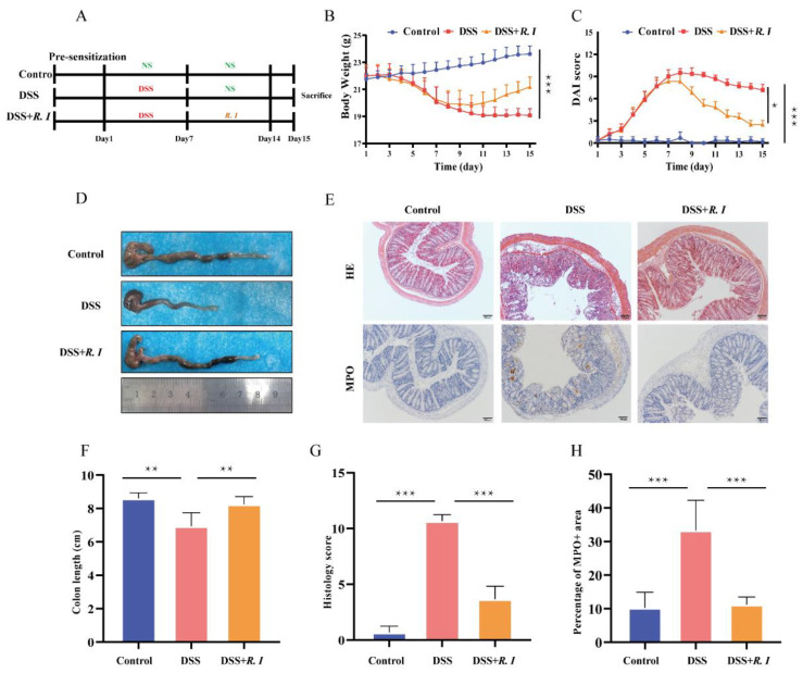
Figure 1.
Mice with DSS-induced intestinal damage and clinical symptoms treated with R. intestinalis. (A) Schematic of the effect of R. intestinalis on DSS-induced colitis in mice. (B) The disease activity index (DAI) scores of the control, DSS, and DSS + R. I treated mice. (C) The body weights of mice in the three groups are plotted and presented as the mean. (D) Illustration of the colons of the mice in each of the three groups. (E) H&E staining for the three groups. Representative images of MPO immunohistochemical staining of colon sections from the three groups. (F) On Day 15, the colon lengths of the three groups were measured. (G) The histology score of H&E staining. (H) The percentage of MPO + area. The statistics are presented as the mean ± SD of three replicates; * p < 0.05, ** p < 0.01, and *** p < 0.001.

