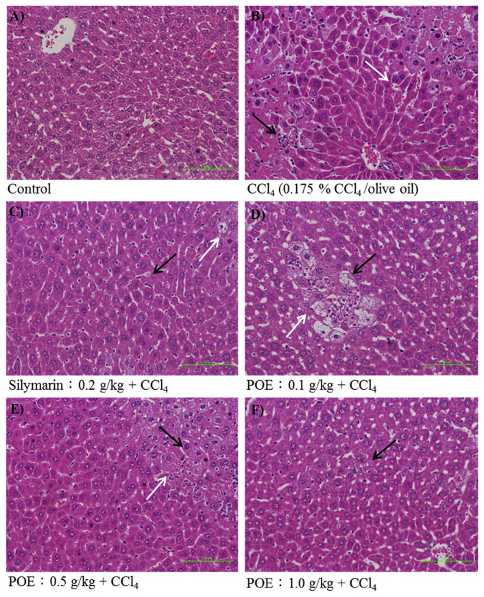
Fig. 5.
The hepatic histological analyses of silymarin (0.2 g/kg) and POE (0.1, 0.5, 1.0 g/kg) on CCl4-induced acute liver injury in mice. Liver tissues were stained with H & E (200×). Cell necrosis was displayed by black arrow and vacuole formation was displayed by white arrow. (A) Control group; (B) animals treated with 0.175% CCl4 (10 mL/kg of bw); (C) animals pre-treated with silymarin (0.2 g/kg) and then treated with CCl4; (D–F) animals pre-treated with POE (0.1, 0.5 and 1.0 g/kg) and then treated with CCl4.

