
Figure 1.
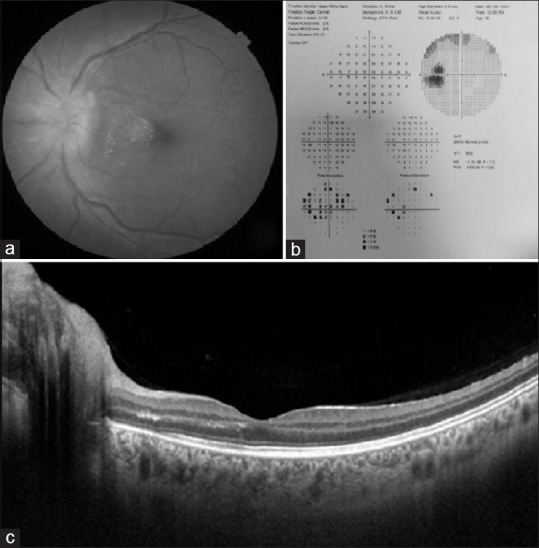
(a) Fundus clinical picture of the patient at presentation; (b) HVF 30-2 of the patient showing an enlargement of the blind spot (c) SS-OCT of the macula showing some exudation in the outer plexiform layer
Official websites use .gov
A
.gov website belongs to an official
government organization in the United States.
Secure .gov websites use HTTPS
A lock (
) or https:// means you've safely
connected to the .gov website. Share sensitive
information only on official, secure websites.
(a) Fundus clinical picture of the patient at presentation; (b) HVF 30-2 of the patient showing an enlargement of the blind spot (c) SS-OCT of the macula showing some exudation in the outer plexiform layer