
Figure 2.
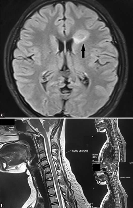
(a) MRI brain and orbit in fat-suppressed FLAIR mode showing a patch of subcortical white matter T2 hyperintensity lesion (marked with black arrow) in the right frontal subcortical area, suggestive of atypical demyelinating pathology. (b) MRI spine with contrast showed T2-weighted lesion between C2 and C7 vertebral levels with swollen cervical cord and edema (marked with white arrow).MRI = magnetic resonance imaging