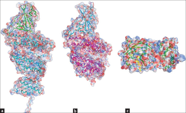
Figure 4.
Molecular docking of severe acute respiratory syndrome coronavirus 2 proteins with receptors/ligands. (a) A cryo-EM structure of severe acute respiratory syndrome coronavirus 2 spike protein RBD (green) in complex with human ACE2 (cyan) (PDB: 6M17 [69]). (b) A crystal structure of severe acute respiratory syndrome coronavirus 2 spike protein RBD (cyan) bound with ACE2 (pink) (PDB: 6M0J [18]). (c) A crystal structure of Mpro (green) in complex with a peptide-like inhibitor N3 (yellow) (PDB: 6 LU7 [77]). Blue and red colors on the sphere presentation of the protein structures indicate the positive and negative charged force fields, respectively

