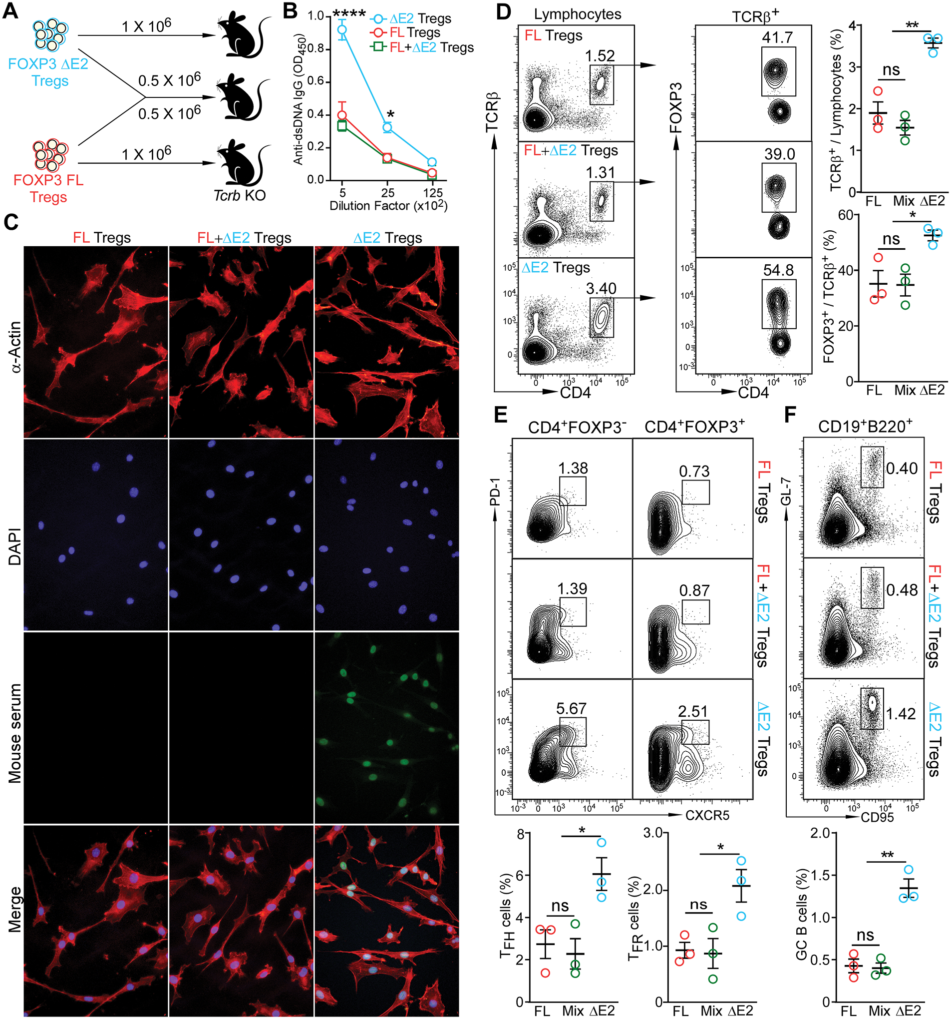
Figure 7. FOXP3 ΔE2 Tregs are sufficient to induce autoantibody and enhanced TFH and GC B cell response.

(A) Experimental design. FOXP3 FL and FOXP3 ΔE2 Tregs were purified by FACS sorting. 1 × 10⁶ purified Tregs were adoptively transferred into Tcrb deficient recipients by tail vail injection. Recipient mice were analyzed 3 months after cell transfer. (B) ELISA quantification of anti-dsDNA IgG in the serum of the recipient mice. (C) Representative images of serum anti-ANA IgG (at 1:80 dilution of serum) in the recipient mice detected with fixed mouse 3T3 fibroblast cells. Mouse serum: sera collected from recipient mice receiving indicated Tregs were used as primary antibody and FITC anti-mouse IgG was used as secondary antibody to detect the existence of anti-ANA IgG in sera. (D) Percentage of donor cells and percentage of donor cells still remaining FOXP3+ in the spleen of recipient mice. (E) TFH (left panels) and TFR (right panels) in the spleen of recipient mice. (F) Germinal center B cells in the spleen of recipient mice. Data represent mean ± SEM (n = 3 mice) from one of 2 independent experiments. ns: not significant; *: p < 0.05; **: p < 0.01; ****: p < 0.0001 by two-way ANOVA (B) or one-way ANOVA (D – F) with Bonferroni post-hoc test.
