
Figure 7. In human aortic endothelial cells (HAECs), mitochondrial DNA copy number, shape, and membrane potential are modulated by sex hormones.
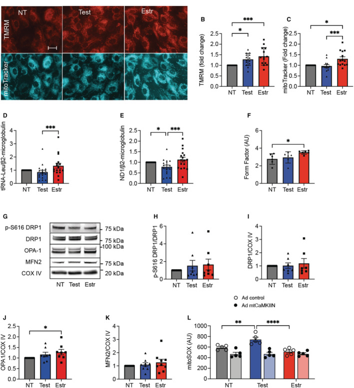
A, Representative images of tetramethylrhodamine methyl ester (TMRM) (mitochondrial membrane potential) and mitoTracker signal in HAECs treated with sex hormones or diluent (nontreated [NT]) for 72 hours. Scale bar=50 μm, ×20. B, Quantification of the TMRM fluorescence. C, Quantification of mitoTracker fluorescence. B and C, Each data point indicates 1 independent experiment (n=14). D and E, Quantitative polymerase chain reaction for genes encoded by mitochondrial DNA normalized to β2‐microglobulin: (D) tRNA‐Leu and (E) for NADH‐ubiquinone oxidoreductase chain 1. n=16 (D), n=17 (E) biological replicates. F, Quantification of mitochondrial form factor. Cells labeled with mitoTracker were analyzed. G through K, Representative immunoblot and quantification for active phosphorylated (p‐)DRP1, DRP1, OPA1, and MFN2 in mitochondrial fractions treated with sex hormones or diluent (NT) for 72 hours. COXIV as loading control. n=8 (H through J) and 9 (K) independent immunoblots. (L) Mitochondrial superoxide levels by mitoSOX fluorescence in HAECs transduced with adenovirus expressing mtCaMKIIN or control and treated with sex hormones or diluent (NT) for 72 hours. n=5 independent experiments. *P<0.05, **P<0.01, ***P<0.005, ****P<0.001 by Kruskal‐Wallis test (F), Friedman test (B through E and H through K), 2‐way ANOVA (L). COXIV indicates cytochrome c oxidase subunit IV; DRP1, dynamin‐related protein 1; Estr, estradiol; MFN2, mitofusin 2; mtCaMKIIN, mitochondrially targeted peptide inhibitor of CaMKII; OPA1, optic atrophy type 1; Test, testosterone.