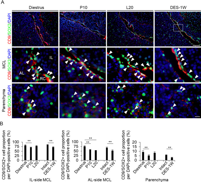
Fig. 2.
Detection of CD9/SOX2- and CD9/PRL-positive cells in the IL-side and AL-side MCLs and AL parenchyma. (A) Double immunohistochemistry for CD9 and SOX2 at diestrus (8–10-week-old female rats (Diestrus)), 10 days of pregnancy (P10), and 20 days of lactation (L20) and after 1 week of DES treatment (DES-1W). Merged image of CD9 and SOX2 immunohistochemistry and DAPI staining are shown (First row: low magnification image of pituitary, second row: magnified view of the MCL, third row: magnified views of the parenchyma). White arrowheads indicate CD9/SOX2-positive cells. (B) The proportion of CD9/SOX2-positive cells per DAPI-positive cells in Diestrus, P10, and L20, DES-untreated rats (Intact), and DES-1W in the IL-side MCL, AL-side MCL, and AL parenchyma (Parenchyma). AL, anterior lobe; IL, intermediate lobe; RC: Rathke’s cleft. Scale bars: 200 μm (first row of A) and 20 μm (second and third rows of A). ** P < 0.01.

