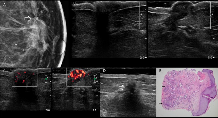
Figure 2.
Images of a 69-year-old woman with bloody left nipple discharge, retraction, and discoloration due to nipple adenoma. Physical examination reported firmness and bluish discoloration of the left nipple. A: Spot magnification mediolateral mammographic image of the left breast demonstrates an area of architectural distortion at the 12-o’clock position in the retroareolar left breast (arrow) with a few associated amorphous calcifications. The left nipple was mammographically unremarkable. B: On US, no discrete mass was seen in either nipple (right on left and left on right). C: Transverse power Doppler image shows hypervascularity of the left nipple (right-hand image) compared to the right. D: Additional transverse sonographic evaluation of the left breast (using a standoff pad) revealed a 15-mm hypoechoic, irregular mass at the 12-o’clock position, 1 cm from the nipple (open arrow), with posterior shadowing, corresponding to the mammographic distortion. US-guided core-needle biopsy showed radial scar with microcalcifications, ductal epithelial hyperplasia, and sclerosing adenosis; associated grade 1 ductal carcinoma in situ was found at excision. E: Histopathology (hematoxylin and eosin, 4x) from punch biopsy of the left nipple shows proliferation of irregular ductal structures (arrows), some of which demonstrate usual ductal hyperplasia, extending to multiple margins of the biopsy, consistent with nipple adenoma.

