Fig. 2.
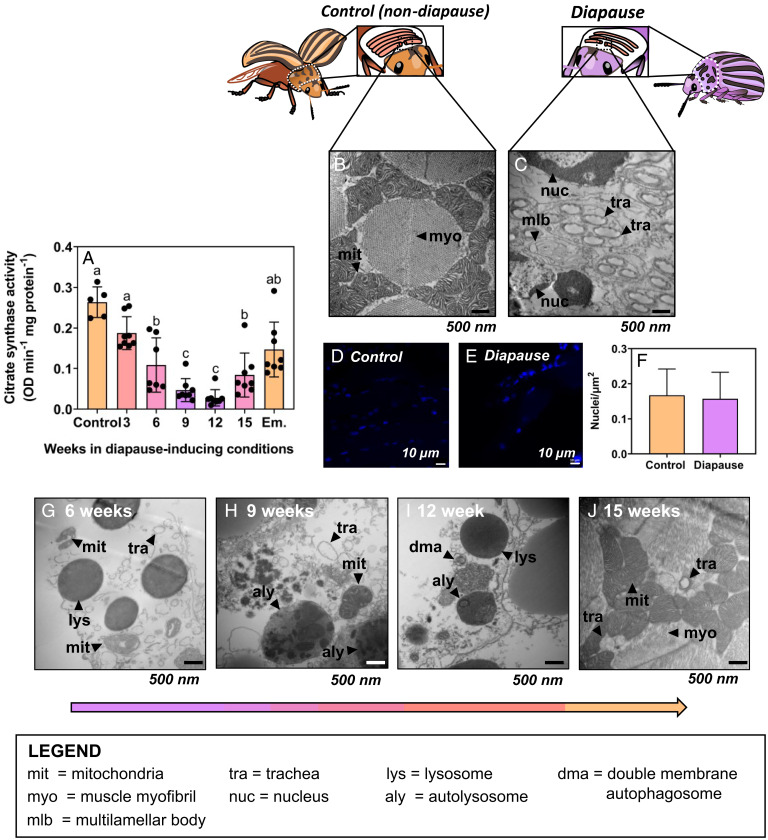
Functional mitochondria are absent from Colorado potato beetle flight muscle during diapause, but beetles reverse this mitochondrial breakdown upon diapause emergence. (A) Mean ± SD citrate synthase activity as a proxy for mitochondrial abundance in flight muscle of beetles as they enter diapause (3–9 wk), during diapause (9–15 wk), and upon emergence (Em). Groups were compared using a 1-way ANCOVA with protein content as a covariate, and different letters denote significant differences among treatments (P < 0.05; SI Appendix, Table S1). (B, C) Representative transmission electron micrographs of flight muscle cross sections from (B) nondiapausing and (C) diapausing beetles (19,000× magnification). Myofibrils (myo) and mitochondria (mit) are mostly absent from diapausing flight muscle; mlb, multilamellar body. (D–F) DAPI staining of flight muscle nuclei in nondiapausing (D) and diapausing (E) beetles indicates no differences in nuclear integrity of flight muscle cells between treatments. (G–J) Representative transmission electron micrographs of flight muscle cross sections from beetles entering diapause (G–I) and emerged beetles (J). Control beetle flight muscle has densely packed mit surrounding large muscle myo, while diapausing beetle flight muscle lack mit but still have nuclei (nuc) and trachea (tra). As beetles enter diapause and during diapause maintenance (6–15 wk; G–I), there are lysosomes (lys), double membrane autophagosomes (dma), and autolysosomes (aly) containing broken-down mit inside, indicating active autophagy in diapausing flight muscle. When beetles emerge from diapause, their flight muscle mitochondrial abundance is recovered (J) and the autophagy machinery (lys, aly, dma) observed during diapause is gone.

