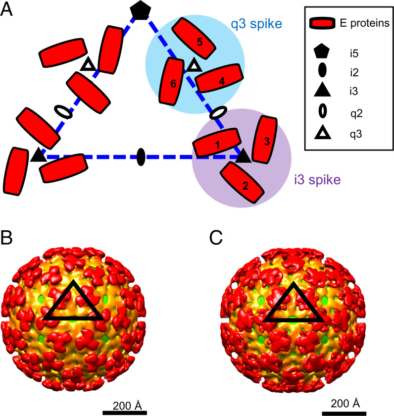
Fig. 1.
Low-pH EEEV cryo-EM reconstructions with icosahedral symmetry imposed. (A) Diagram of alphavirus E1–E2 glycoproteins arranged with T = 4 icosahedral symmetry. The dashed triangle (blue) defines an asymmetric unit in an alphavirus structure with T = 4 icosahedral symmetry. Quasitwofold (q2), q3, icosahedral twofold (i2), i3, and icosahedral fivefold (i5) symmetry elements are labeled. The envelope glycoproteins (E proteins) containing E1 and E2 are shown in red. One asymmetric unit contains one i3 E2–E1 protein (labeled 1) and three q3 E2–E1 proteins (labeled 4, 5, and 6). (B) Cryo-EM reconstruction of prefusion state 1 of EEEV. (C) Cryo-EM reconstruction of prefusion state 2 of EEEV. The black triangles indicate an asymmetric unit of the EEEV cryo-EM reconstruction with T = 4 icosahedral symmetry. The cryo-EM maps are radially colored. The virus membrane is shown in green (∼240 Å from the virus center), and the glycoprotein shell is depicted in orange (∼250 Å from the virus center) and red (∼350 Å from the virus center).

