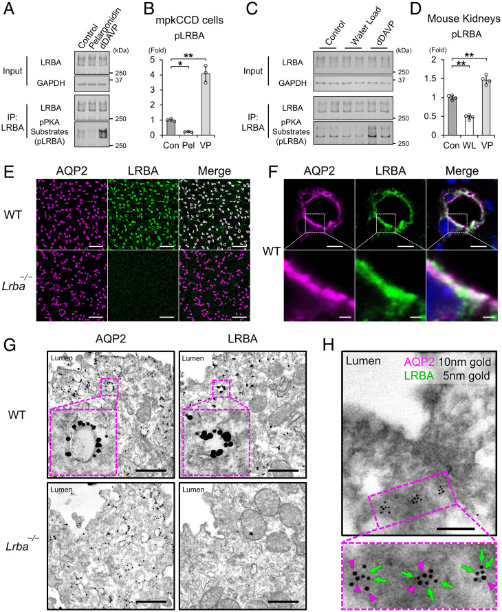
Fig. 2.
LRBA colocalized with AQP2 at vesicles in the subapical region. (A and B) The effects of pelargonidin and vasopressin on LRBA phosphorylation in mpkCCD cells (n = 3). (C and D). The effects of water load and vasopressin on LRBA phosphorylation in vivo (n = 4). (E and F) Immunofluorescence staining showing that AQP2 (magenta) and LRBA (green) are colocalized in the kidneys of WT mice. (E) Scale bars, 100 μm. (F) Scale bars: top, 5 μm; bottom, 1 μm. (G) Immuno-electron microscopy showing that AQP2 and LRBA are localized at vesicles in renal collecting ducts of WT mice. Scale bars, 500 nm. (H) Colocalization of AQP2 and LRBA at the same intracellular vesicles. Double-label immuno-electron microscopy of AQP2 (10-nm gold indicated by magenta arrowheads) and LRBA (5-nm gold indicated by green arrows) in renal collecting ducts of WT mice. Scale bars, 200 nm. Data are reported as mean ± SD. *P < 0.05, **p < 0.01 by Dunnett's test. IP, immunoprecipitation; Pel, pelargonidin; VP, dDAVP; WL, water load.

