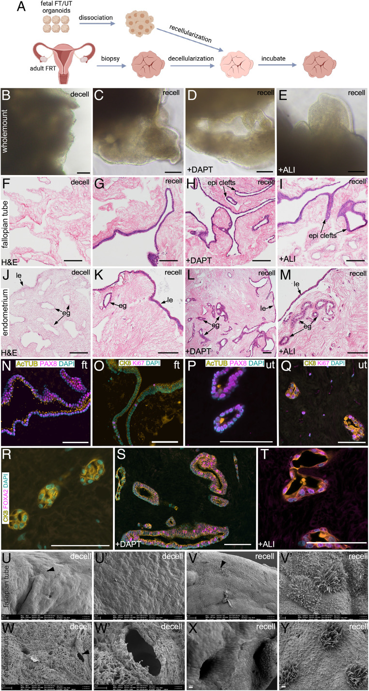
Fig. 5.
Transplantation of human fetal organoids onto decellularized adult tissue scaffolds leads to epithelial regeneration. (A) Schematic showing the experimental schedule used in B–Y. FRT, female reproductive tract. (B–E) Representative whole-mount bright-field images of decellularized (decell) (B) and recellularized (recell) (C–E) adult tissue scaffolds cultured in the presence of vehicle (C) or DAPT (D) or at an ALI (E) (n = 3 per group). (F–M) Representative histological images of adult FT (F–I) and uterine (J–M) tissue-derived scaffolds before (F and J) and after (G–I and K–M) fetal cell transplantation. Scaffolds were cultured in the presence of vehicle (G and K) or DAPT (H and L) or at an ALI (I and M) (n = 3 per group × 10 tissue sections of 5-μm thickness were examined per replicate). (N–Q) Coimmunostaining for PAX8 (secretory cells) and AcTUB (ciliated cells) (N and P) and Ki67 (proliferating cells) and CK8 (epithelial cells) (O and Q) in recellularized FT (N and O) and uterine (P and Q) scaffolds. (R–T) Coimmunostaining for FOXA2 and CK8 in recellularized uterine scaffolds cultured in the presence of vehicle (R) or DAPT (S) or at an ALI (T); n = 3 biological replicates per group × 5 tissue sections of 5-μm thickness were stained per replicate. Scanning electron microscopy of decellularized (U, U′, W, and W′) and recellularized (V, V′, X, and Y) FT (U, U′, V, and V′) and uterine (W, W′, X, and Y) scaffolds (n = 3 per group). The epithelial cell layer consisting of an admix of ciliated and secretory cells was present after but not before recellularization. U′, V′, and W′ represent high-magnification images of areas marked by arrowheads in U, V, and W, respectively. Epi clefts, epithelial clefts. (Scale bars, 100 μm unless indicated otherwise.)

