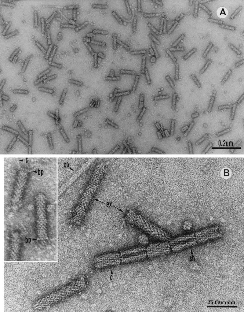
FIG. 1.
Electron micrographs of enterocoliticin particles after negative staining. (A) Extended and contracted phage tail-like structures. (B) Higher magnification of enterocoliticin particles with different structural elements, showing the similarity to the architecture of phage tails (ex, extended particles; c, contracted particles; sh, sheath; co, core). (Inset) Particles with base plate (bp) and spikes (s).

