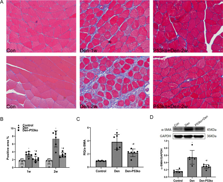
Fig. 4.
P53 KO attenuated muscle fibrosis after d enervation. A, B Masson’s trichrome staining of the muscle tissue and quantification of the positive area as percentages of the fiber area (blue collagen fiber), showing that muscle fibrosis was reduced when p53 was knocked out. Scale bar, 50 μm. *P<0.05 versus control; #P<0.05 versus Den (n=6/group). C Elevated expression of α-SMA mRNA in both the WT and the P53 KO groups of mice after denervation was determined by qPCR. Level of elevation of α-SMA in the WT group of mice was much higher than the P53 KO group after denervation. *P<0.05 versus control; #P<0.05 versus Den (n=6/group). D Levels of α-SMA protein determined by western blotting showed a trend similar to that of the mRNA expression. *P<0.05 versus control; #P<0.05 versus Den (n=6/group). WT, wild type; KO, knockout

