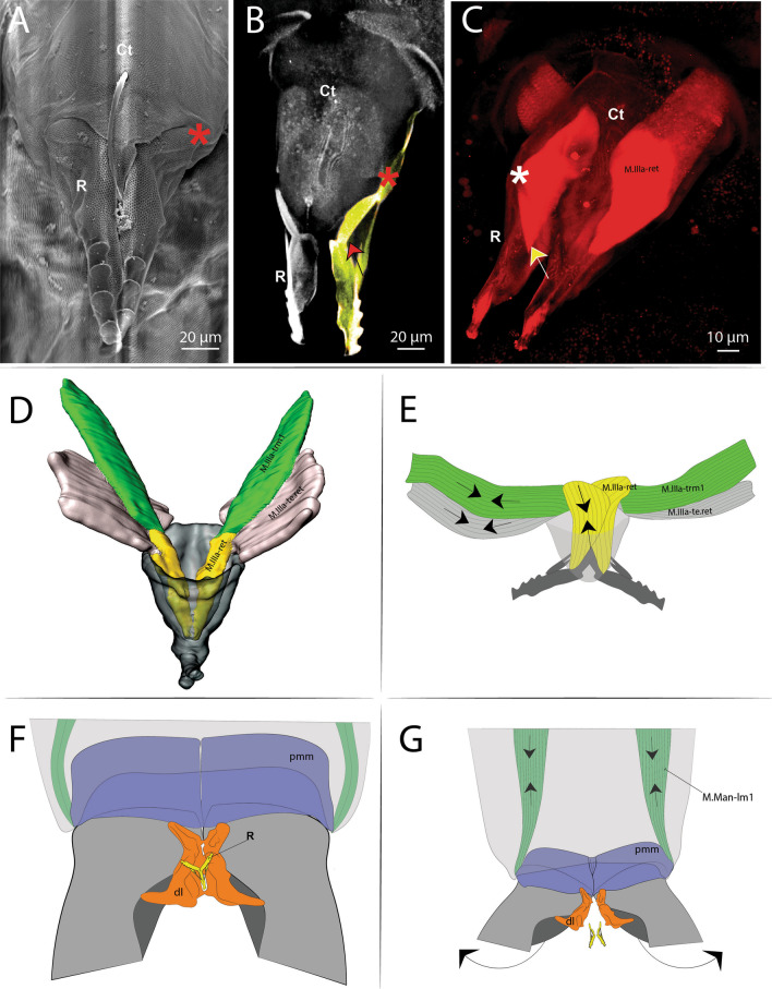
Fig. 16.
Morphology and function of the retinaculum. A Scanning electron microscopy (SEM) image (anterior view) of the retinaculum; B Confocal microscopy (cLSM) image at 405 nm (view from anterior) of the retinaculum; yellow marking showing the Rami; red arrow showing the point of connection of the M.IIIa-ret muscle internally to the rami; C Confocal microscopy (cLSM) image at 555 nm (stained with phalloidin) (view from anterior) of the retinaculum; yellow arrow showing the point of connection of the M.IIIa-ret muscle internally to the rami; D Morphological reconstruction using micro computer tomography (MicroCT) (view from posterior) of the retinaculum; E Schematic representation showing the muscular function of the retinaculum; F, G Schematic representation showing a hypothesis on how the furca may be released from the retinaculum. Ct: corpus tenaculi; R: retinaculum ramus or rami; DL: dens lock; The “*” means the Pivot point of articulation between Rami and Corpus tenaculi

