Fig. 3.
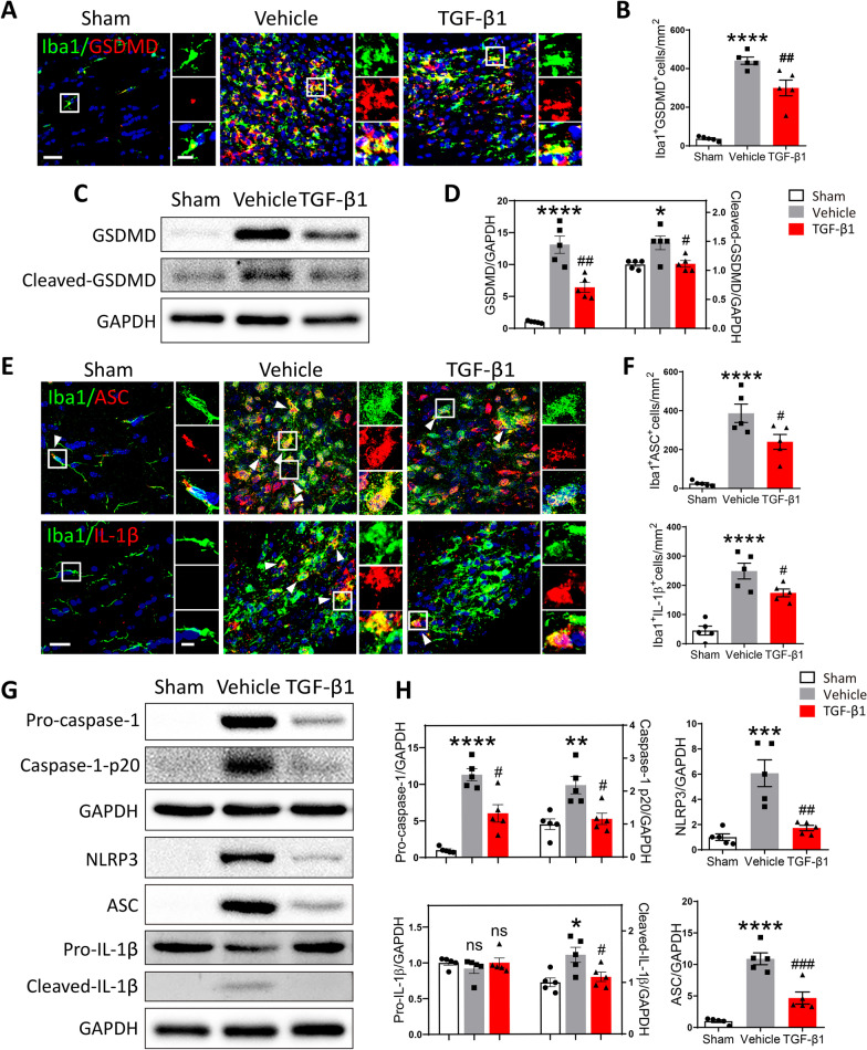
Preventative administration of TGF-β1 attenuated the pyroptosis of microglia in LPC-modeling mice. A Representative confocal images showing Iba1 and GSDMD in the corpus callosum among different groups, scale bar = 30 μm. Local enlarged views were presented, scale bar = 10 μm. B Quantitative analysis of the number of Iba1+GSDMD+cells was performed using One-way ANOVA followed by Tukey’s multiple comparisons test. ****P < 0.0001 versus Sham mice; ##P < 0.01 versus Vehicle mice. N = 5 per group. C The protein expression of GSDMD and Cleaved-GSDMD in the corpus callosum among three group was detected by Western blot. D Quantitative analysis of Western blot was performed using One-way ANOVA followed by Tukey’s multiple comparisons test. *P < 0.05, ****P < 0.0001 versus Sham group; #P < 0.05, ##P < 0.01 versus Vehicle group. N = 5 per group. E ) Representative confocal images showing Iba1 with ASC or IL-1β in the corpus callosum among different groups, scale bar = 20 μm. Local enlarged views were presented, scale bar = 5 μm. White arrowhead pointed to the immunofluorescent double-labeling cells. F Quantitative analyses of the number of Iba1+ASC+cells and Iba1+IL-1β+cells were performed using One-way ANOVA followed by Tukey’s multiple comparisons test. ****P < 0.0001 versus Sham mice; #P < 0.05 versus Vehicle mice. N = 5 per group. G The protein expression of Pro-caspase-1, Caspase-1-p20, NLRP3, ASC, IL-1β, and Cleaved-IL-1β in the corpus callosum among three groups was detected by Western blot. H Quantitative analysis of Western blot was performed using One-way ANOVA followed by Tukey’s multiple comparisons test. *P < 0.05, **P < 0.01, ***P < 0.001, ****P < 0.0001, n.s. no significance versus Sham group; #P < 0.05, ##P < 0.01, ###P < 0.001, n.s. no significance versus Vehicle group. N = 5 per group

