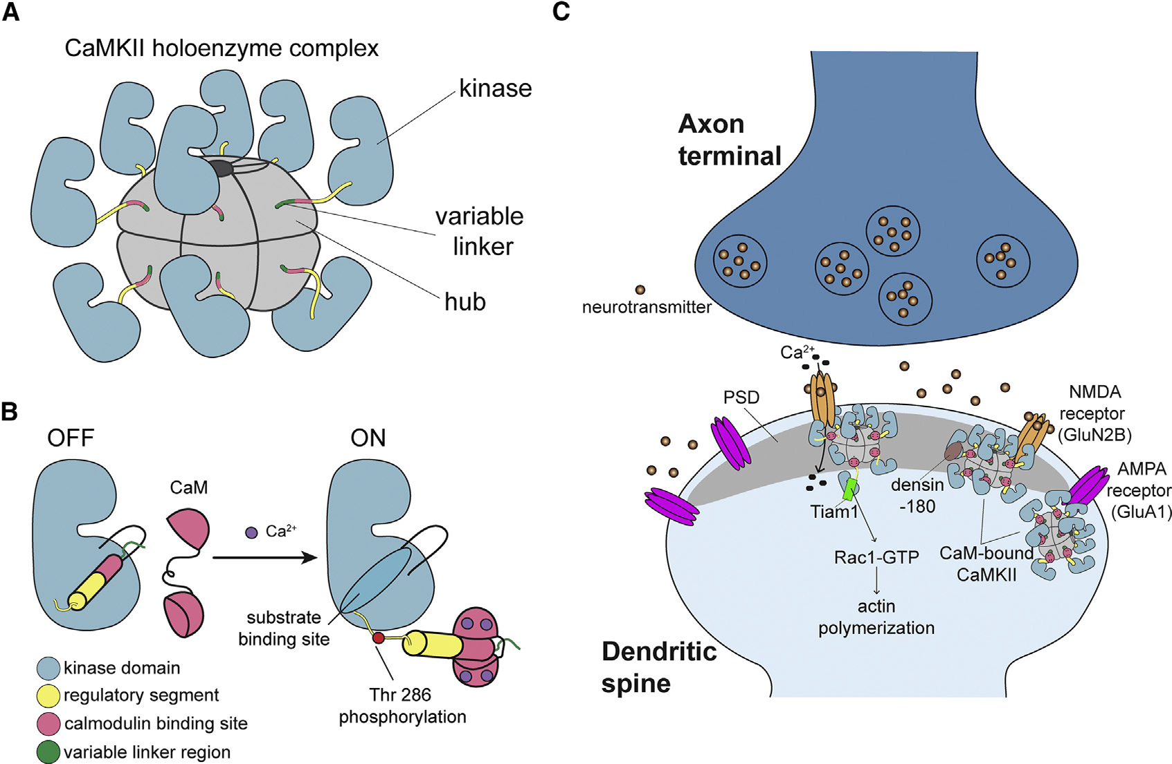
Figure 1. CaMKII architecture and the interaction partners at excitatory synapses.

(A) The architecture of a dodecameric CaMKII holoenzyme.
(B) Ca2+/CaM binding activates CaMKII by competitively binding the regulatory segment, thereby freeing the substrate binding site. Active CaMKII autophosphorylates at Thr 286.
(C) CaMKII interactions at the excitatory postsynaptic structure, mostly in the postsynaptic density (PSD), of the dendritic spine.
