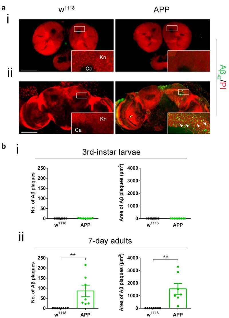
Figure 1.

Diffuse amyloid deposits are abundant in the mushroom body (MB) in seven-day-old APP adults but not in third-instar APP larvae. (a) Representative images. Aβ deposits were stained with anti-Aβ42 antibody (green). Nuclei were stained with PI (red). The Kenyon (Kn) cell region (boxed) was zoomed in to display Kn cells and Aβ deposits. (i) Immunostaining of brains of third-instar larvae shows a negligible Aβ42 signal in APP flies compared to no Aβ42 signal in w1118 flies. (ii) Immunostaining of brains of seven-day-old adults shows evident Aβ deposits in APP flies compared to w1118 flies. Arrowheads indicate Aβ deposits. No Aβ42 signal was detected in the Calyx (Ca) region. Scale bar represents 100 μm. (b) Aβ deposits were quantified by both number and size. (i) Quantification of Aβ deposit numbers and areas in the third-instar larval brain Kn region. n = 9 ~ 10. (ii) Quantification of Aβ deposit numbers and area in the seven-day-old adult brain Kn region. n = 8 ~ 9. **p < 0.01; unpaired student’s t-test. All data are shown as mean ± SEM.
