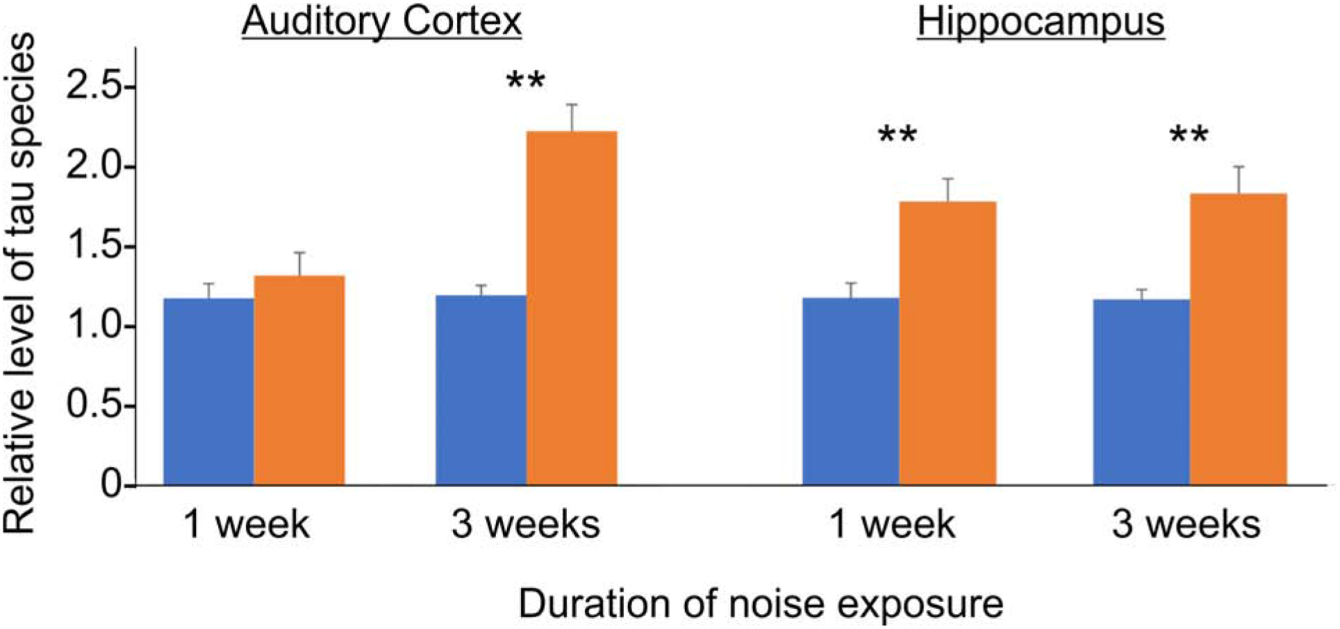
Figure 3:

Density analysis taken from Western blots of hippocampal tissue probed with an antibody against the phosphorylation site at Serine 396 of tau. Levels were normalized to the tau levels in the control group and done with three replicates. Error bars represent the standard deviation. **p<0.001 compared to control. Reproduced with permission from (Cheng et al., 2016).
