Figure 4.
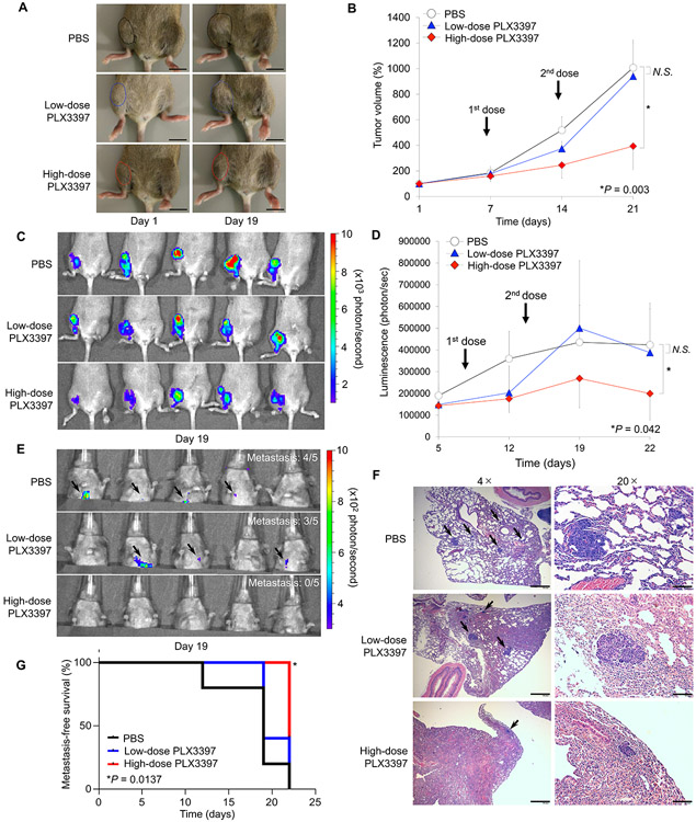
CSF-1R inhibition by PLX3397 blocks tumor growth and improves metastasis-free survival in orthotopic osteosarcoma mouse model. A. Macroscopic appearance of LM8-Luc tumors in C3H/JeJ mice on Days 0 and 19 after tumor cell inoculation. Mice were inoculated intratibially with LM8-Luc cells (1 × 10⁶ cells/site) and were systemically treated with control PBS, low-dose PLX3397 (5 mg/kg), or high-dose PLX3397 (10 mg/kg) on Day seven and 14. Tumor masses are outlined by a dotted line. B. Tumor growth in an orthotopic LM8 osteosarcoma xenograft model of each treatment group (n = 5 per group). Data was expressed as mean tumor volume ± SD. *p < 0.05, as compared with high-dose PLX3397 and PBS group; one-way ANOVA corrected for multiple comparisons. C. Luminescence intensity from the primary tumors of each treatment group measured on Day 19 using an IVIS. D. Monitoring of luminescence intensity from the primary tumors of each treatment group. Data was expressed as mean tumor volume ± SD. *p < 0.05, as compared with high-dose PLX3397 and PBS group; one-way ANOVA corrected for multiple comparisons. E. Lung metastases measured on Day 19 using an IVIS. F. Lung metastases validated by H&E staining. Black arrow represents metastatic foci in the lung. Scale bar, 200 μm (left). Scale bar, 40 μm (right). G. Kaplan-Meier curves showing metastasis-free survival for each group of mice. Log-rank test was performed between PBS control group (black line) and low-dose PLX3397 group (blue line; p = 0.353) or high-dose PLX3397 group (red line; *p = 0.0014).

