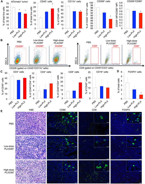
Figure 5.
Systemic treatment of PLX3397 depletes TAMs and increases lymphocyte infiltration into LM8 osteosarcoma. A. Composition of tumor cells and TAMs evaluated by flow-cytometric analysis using the dissociated LM8 tumor cells. Data were represented as mean ± SEM; n = 3; Mann–Whitney test: *p < 0.05. B. Flow cytometry analysis of TAMs (CD45+CD11b+CD206+; left) and CD8 T cells (CD45+CD3+CD8+; right) within the dissociated LM8 tumor cells and representative flow data. C. Composition of infiltrating immune cells (CD3+, CD4+, and CD8+ cells) evaluated by flow-cytometric analysis using the dissociated LM8 tumor cells. Data were represented as mean ± SEM; n = 3; Mann – Whitney test: *p < 0.05. D. Composition of infiltrating FOXP3+ regulatory T cells evaluated by immunohistochemistry using the dissociated LM8 tumor cells. Data were represented as mean ± SEM; n = 3; Mann–Whitney test: *p < 0.05. E. Distribution of infiltrating CD68+ macrophages, CD8+ T cells, and FOXP3+ regulatory T cells in PLX3397- or PBS-treated tumors (green). Nuclei were stained with DAPI (blue). Left, H&E staining images. Scale bars, 50 μm (lower) and 10 μm (upper).

