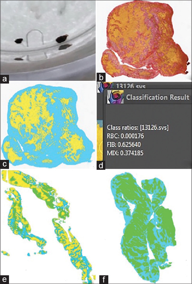
Figure 1.

(a) A study subject clot sample prior to sectioning. (b) Clot sample with MSB stain. (c) Clot sample after segmentation. RBCs were highlighted green, mature and fresh fibrin were grouped and highlighted blue, and “mixed” area was highlighted yellow. (d) Quantification of clot sample in panel C showing it is fibrin rich. (e) Example of mixed-type clot. (f) Example of RBC-rich clot. RBC: Red blood cells, MSB Martius Scarlett Blue
