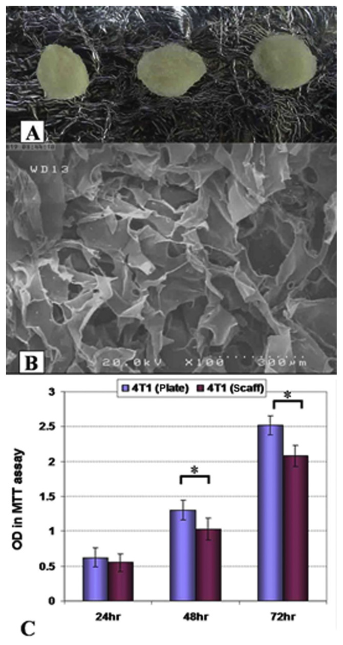
Figure 1.
(A) Macroscopic and (B) electronic microscopy structure of collagen–chitosan scaffolds. (C) 4T1 tumor growth in three-dimensional (3D) and two-dimensional conditions. Scaffolds presented an appropriate porosity for cell infiltrations. Cultured 4T1 cells showed lower growth rates on 3D scaffolds surfaces after 48 hours and 72 hours of culture (p < 0.05). OD = Optical Density; MTT = 3-(4,5-dimethylthiazol-2-yl)-2,5-diphenyltetrazolium bromide; scaff = scaffold, *: p < 0.05.

