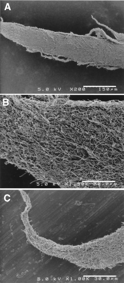
FIG. 3.
Scanning electron micrographs of spine-like projections sticking out of the fluffy granules (sea urchin granules) in reactor II. (A) Whole view of a projection (bar, 150 μm). (B) Higher magnification of the bottom part of the projection (bar, 20 μm). (C) Higher magnification of the tip of the projection (bar, 30 μm)

