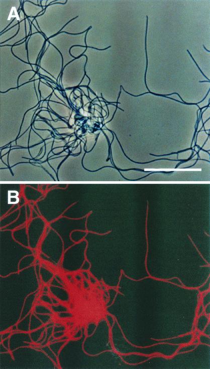
FIG. 5.
In situ hybridization of strain UNI-1 cells isolated in this study. The cells were hybridized with the rhodamine-labeled GNSB633 probe. (A) Phase-contrast micrograph of strain UNI-1 cells (bar, 10 μm). (B) Fluorescent micrograph of the same field as panel A, showing that all cells were stained with the thermophilic UASB cluster-specific probe GNSB633.

