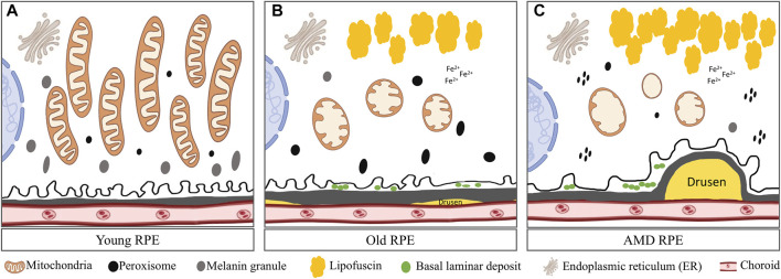
FIGURE 2.
Comparison of mitochondria in young, aged and AMD RPE: (A) Young RPE cell contains numerous mitochondria with long axes, usually oriented from the apical to the basal surfaces of the RPE and are parallel to one another. The mitochondrial cristae are well preserved. Several peroxisomes appeared as small, round, electron-dense organelles. Plenty of melanin granules exist in the cells. (B) In aged RPE cell, mitochondria show membrane disorganization and loss of cristae. Accumulated lipofuscin presents in the cell. Several peroxisomes of various density, shape and size were distributed randomly in the cytoplasm. Less melanin granules appear in the cell. Also, small drusen forms underneath BrM and basal lamina deposits forms in between the cell and BrM. (C) In AMD RPE, advanced mitochondrial alterations occur. Most mitochondria had severe disorganization of membranes that varied from focal to complete loss of cristae. Peroxisomes are clustered and aggregated in the cell. Large and soft drusen forms underneath the BrM.

