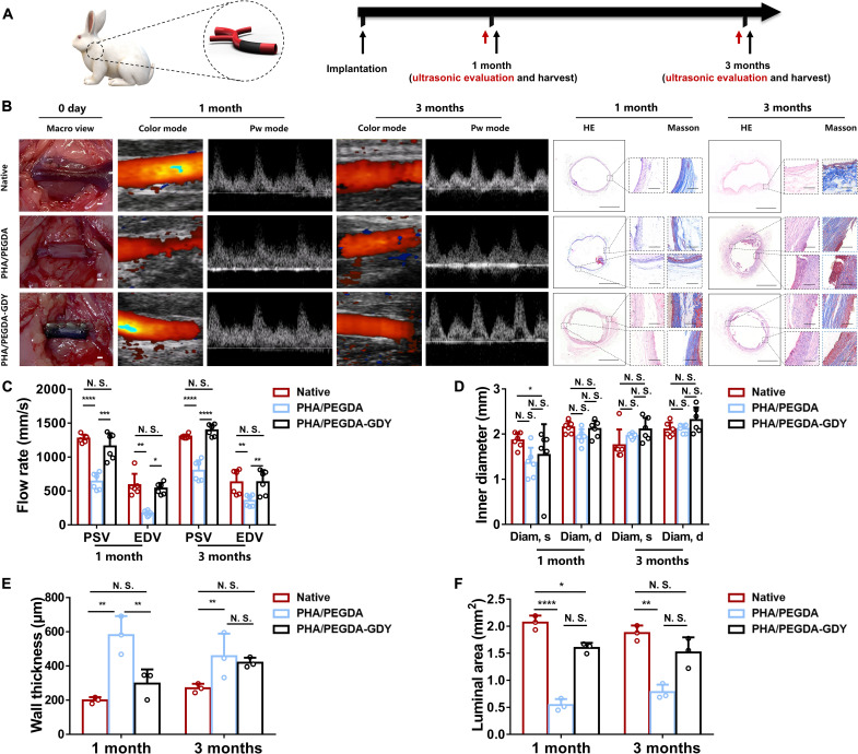
Fig. 7. In vivo implantation of gel vascular grafts.
(A) Gel vascular grafts were implanted in rabbit model of carotid artery defect, and then ultrasound and histological evaluation of the vascular grafts were performed after 1 and 3 months of implantation. (B) Macroscopic views upon implantation, ultrasound images, and HE and Masson staining images after implantation of vascular grafts of gels. Scale bars, 1 mm (macro view), 1 mm (panorama), and 200 μm (magnified view). (C) Peak rate of blood flow and (D) inner diameters of vascular grafts of gels at different time points after implantation from ultrasonic evaluation (n = 6). (E) Wall thickness and (F) luminal area of vascular grafts explanted at different time points after implantation from histological analysis (n = 3). ****P < 0.0001, ***P < 0.001, **P < 0.01, and *P < 0.05 by one-way ANOVA with Tukey correction.

