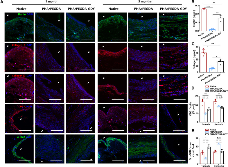
Fig. 8. Immunofluorescence staining of implanted vascular grafts after 1 and 3 months of implantation.
(A) Immunofluorescence staining of elastin (green), collagen I (red), collagen III (red), CD31 (red), and α-SMA (green) of the grafts. White arrows indicate luminal areas. Scale bars, 200 μm. (B) Elastin and (C) collagen content in grafts explanted at the time point of 3 months (n = 3). Quantification analysis of CD31+ cells (D) and percent of α-SMA+ area (E) in vascular grafts (n = 3). ****P < 0.0001, ***P < 0.001, **P < 0.01, and *P < 0.05 by two-way ANOVA with Tukey correction.

