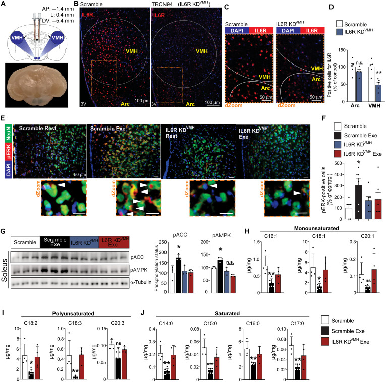
Fig. 5. Hypothalamic IL6 controls muscle fatty acid oxidation after exercise in mice.
(A) Illustration for VMH microinjections and a representative picture of a coronal view of a mouse brain, demonstrating the blue dye staining confirming the anatomical localization of bilateral microinjections in VMH (bottom). KD, knockdown. (B) IL6R (red) in the arcuate nucleus and VMH 7 days after VMH lentivirus injection (n = 5 to 6). Scale bars, 100 μm. (C) dZoom from the orange rectangles highlighted in (B) shows the presence of IL6R (red). Scale bars, 50 μm. DAPI, 4′,6-diamidino-2-phenylindole. (D) IL6R-positive cells in the arcuate and ventromedial nuclei (n = 5 to 6, **P < 0.01 versus scramble). (E) Top panels: Colocalization of ERK1/2 phosphorylation (red) in neurons (green) of VMH (n = 3 to 5). Scale bars, 50 μm. Bottom panels: dZoom from the orange rectangles highlighted in the top panels shows the presence of pERK1/2 in neuronal cells. Scale bars, 10 μm. (F) Positive cells for pERK1/2 in neuronal and nonneuronal cells in VMH of mice (n = 6, *P < 0.05 versus scramble at rest). (G) ACC and AMPK phosphorylation in the soleus muscle (n = 3, *P < 0.05 versus scramble at rest). (H to J) Determination of fatty acid fractions in the gastrocnemius muscle (n = 4 to 6). Unpaired t test was used in (D) and (F). One-way ANOVA was used for statistical analysis in (G) and (H).

