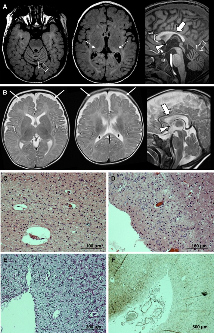
Figure 1.
Brain MRI and histology. (A and B) Relevant neuroimaging features associated with ADAM22 variants, including cerebral atrophy with enlargement of the CSF spaces (thin arrows) and lateral ventricles (asterisks), cerebellar atrophy with prevalent vermian involvement (empty arrows), corpus callosum hypoplasia/thinning (thick arrows) and anterior commissure hypoplasia (arrowheads). Additional diffuse hyperintensity of the supratentorial white matter with bilateral pulvinar involvement (dotted arrows) was noted in one subject on FLAIR images (A) from Patient P4 and (B) from Patient P5. (C–F) Post-mortem examination of brain tissue obtained from Patient P10 (deceased at the age of 28 years). (C) Haematoxylin and eosin-staining (×200 magnification) of the visual cortex, which showed profound atrophy and neuronal depletion with only some pyramidal cells in layers V–VI. (D) Haematoxylin and eosin-staining (×200 magnification) of the medial thalamus which was extremely atrophic and gliotic. (E) PAS staining (×100 magnification) of the frontal cortex which was very atrophic with a vast number of corpora amylacea. (F) Neurofilament SMI32 staining by immunohistochemistry (×40 magnification), showing the pronounced loss of neurons at the sulcal region.

