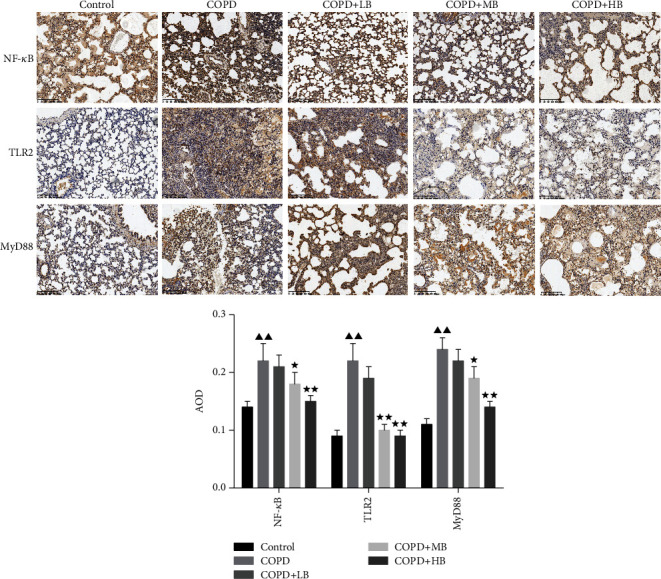
Figure 6.

BA changed the levels of NF-κB, TLR2, and MYD88 in the lung tissue of COPD rats by immunohistochemical staining and semiquantitative analysis (×100). Data were expressed as mean ± SD, n = 3. Compared with the control group, ▲P < 0.05 and ▲▲P < 0.01; compared with the COPD group, ★P < 0.05 and ★★P < 0.01.
