Fig. 5.
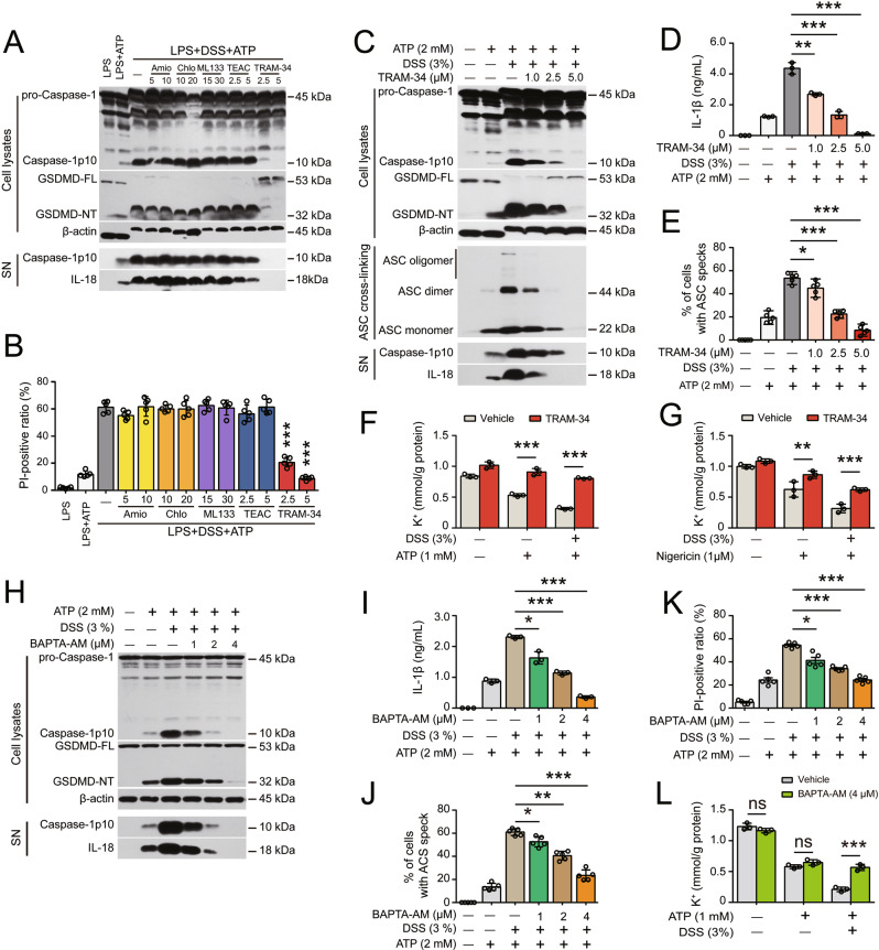
Blocking the KCa3.1 channel attenuates the DSS-induced increase in NLRP3 inflammasome activation by suppressing K+ efflux. After being primed with LPS for 4 h, BMDMs were treated with DSS for 1 h and then stimulated with ATP (30 min). K+ channel inhibitors were added and incubated as follows: TRAM-34 and tetraethylammonium chloride (TEAC) were added before LPS priming for 1 h; and amiodarone (Amio), chlorpromazine (Chlo) and ML133 were added after LPS priming but before DSS for 1 h. The indicated proteins in cell lysates and culture supernatants (SN) were assayed by Western blotting. ASC oligomerization was determined by using ASC crosslinking and immunofluorescence staining. Lytic cell death was determined using PI/Hoechst 33342 staining, and the ratios of PI-positive cells in 5 randomly chosen fields were quantified. IL-1β levels in supernatants were measured with a CBA. The K+ concentration in the cell lysates was measured by a potassium (K+) turbidimetric assay and normalized to the protein concentration in the homogenate (mM/g protein). A, B The effects of several selected K+ channel inhibitors on DSS-mediated promotion of ATP-induced inflammasome activation (A) and lytic cell death (B). C–E TRAM-34 dose-dependently attenuated the DSS-induced increase in ATP-induced inflammasome activation (C), IL-1β secretion (D) and ASC speck formation (E). After sequential treatment with TRAM-34, LPS and DSS, BMDMs were stimulated with ATP (30 min) to activate the NLRP3 inflammasome. F, G TRAM-34 treatment reversed the DSS-induced enhancement of ATP (F)- and nigericin (G)-induced K+ efflux. K+ efflux was induced by ATP (1 mM, 10 min) and nigericin (1 µM, 10 min). H–L Blocking intracellular Ca2+ signaling with BAPTA-AM abrogated the DSS-induced increases in ATP-induced inflammasome activation (H), IL-1β secretion (I), ASC speck formation (J), pyroptosis (K) and K+ efflux (L). After LPS priming and DSS treatment, BMDMs were incubated with BAPTA-AM in Ca2+-free buffer for 40 min prior to ATP stimulation. The data are expressed as the mean ± SD. *p < 0.05, **p < 0.01, ***p < 0.001

