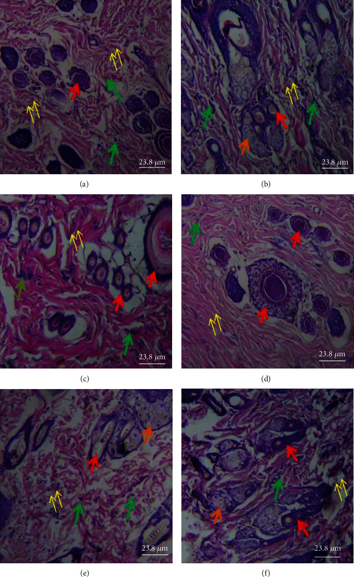
Figure 6.

Histological section of the dermis showing pilosebaceous unit components [hair follicles (red arrow), sebaceous glands (orange arrows)], granulation tissue (yellow arrows), and inflammatory cells (green arrows). (a) Several small hair follicles but no sebaceous glands and loose granulation tissue with moderate inflammatory cells infiltration. (b) Variable-sized hair follicles, large sebaceous glands, and loose granulation tissue with few inflammatory cell infiltrations. (c) Several small hair follicles with perifollicular vasodilatation, no sebaceous glands, loose granulation tissue with eosinophilic collagen fibers, and moderate inflammatory cell infiltration. (d) Several hair follicles of variable sizes and with perifollicular vasodilatation, no sebaceous glands, and fibrotic granular tissue with no obvious inflammatory cell infiltration. (e) Less basophilic hair follicles, few sebaceous glands, and very loose granulation tissue with moderate inflammation cell infiltration. (f) Several small hair follicles and numerous sebaceous glands and loose granulation tissue with moderate inflammatory cell infiltration. Magnification: ×400. (a) Model, (b) vehicle, (c) 1% SSD, (d) 0.3% PCFHE, (e) 1% PCFHE, and (f) 3% PCFHE. PCFHE: P. clappertoniana fruit husk extract; SSD: silver sulfadiazine.
