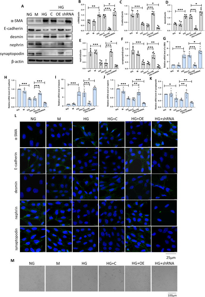
Fig. 2. Sestrin2 attenuates podocyte phenotypic alterations induced by HG.
A–F A representative Western blot and relevant quantification of α-SMA, E-cadherin, desmin, nephrin, and synaptopodin in mouse podocytes (n = 5). G–K The mRNA levels of α-SMA, E-cadherin, desmin, nephrin, and synaptopodin were analyzed by RT-qPCR in mouse podocytes (n = 5). L Representative immunofluorescence images of α-SMA, E-cadherin, desmin, nephrin, and synaptopodin in mouse podocytes, DAPI is a dye for nucleus. M Morphological changes of podocytes cultured under different conditions were analyzed by the inverted microscope. NG: 5.6 mM D-glucose; M: 5.6 mM d-glucose+24.4 mM mannitol; HG: 30 mM d-glucose; HG + C: HG + control shRNA plasmid; HG + OE: HG + Sestrin2 pcDNA; HG + shRNA: HG + Sestrin2 shRNA plasmid. Data are expressed as mean ± SD. *P < 0.05, **P < 0.01, ***P < 0.001. α-SMA α-smooth muscle actin, mRNA messenger RNA, RT-qPCR quantitative real-time polymerase chain reaction.

