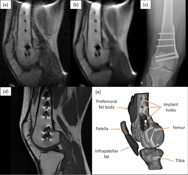
Figure 4.
Images of fixation metallic implant attached to the femur, consisting of a plate and seven screws: (a) sagittal view of a raw low-field image acquired with the 72 mT system (9 mm slice from -weighted 3D-RARE acquisition with in-plane resolution of mm, 12 min scan time, eight years after femoral shaft osteotomy); (b) same, but BM4D-filtered27 and rescaled by to increase the number of pixels; (c) lateral X-ray computed radiography (two weeks after surgery); (d) sagittal view of the same knee, acquired with a Siemens Skyra 3 T system (-weighted 2D-RARE acquisition with slice thickness 3.9 mm and pixel resolution mm, one year after surgery); and (e) 3D reconstruction from -weighted 3D-RARE acquisition with isotropic resolution of 2 mm, 20 min scan time, where selected muscle and fat segments have been removed (eight years after surgery).

