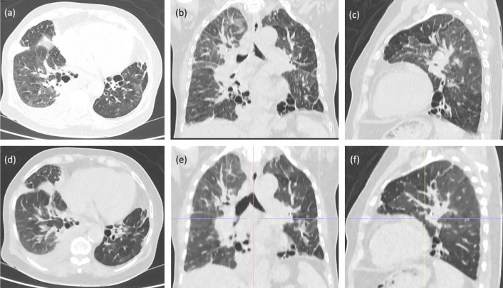
Figure 1.
These figures show chest CT images of a 45-year-old female COVID-19 patient. Top row images show consolidation and crazy-paving pattern in (a) axial, (b) coronal, and (c) sagittal views using the RD-CT protocol. Bottom row images show the same features in (d) axial, (e) coronal, and (f) sagittal views using the ULD-CT protocol with 94% dose reduction.

