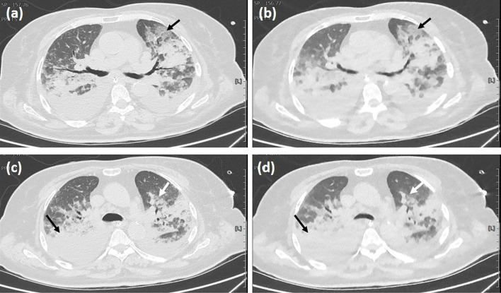
Figure 2.
The axial chest CT images of a 71-year-old female COVID-19 patient are shown. Crazy-paving pattern (black arrow) is shown using the (a) RD-CT and (b) ULD-CT protocols. Ground-glass opacity (white arrow) and consolidation (black arrow) are shown using the (c) RD-CT and (d) ULD-CT protocols.

