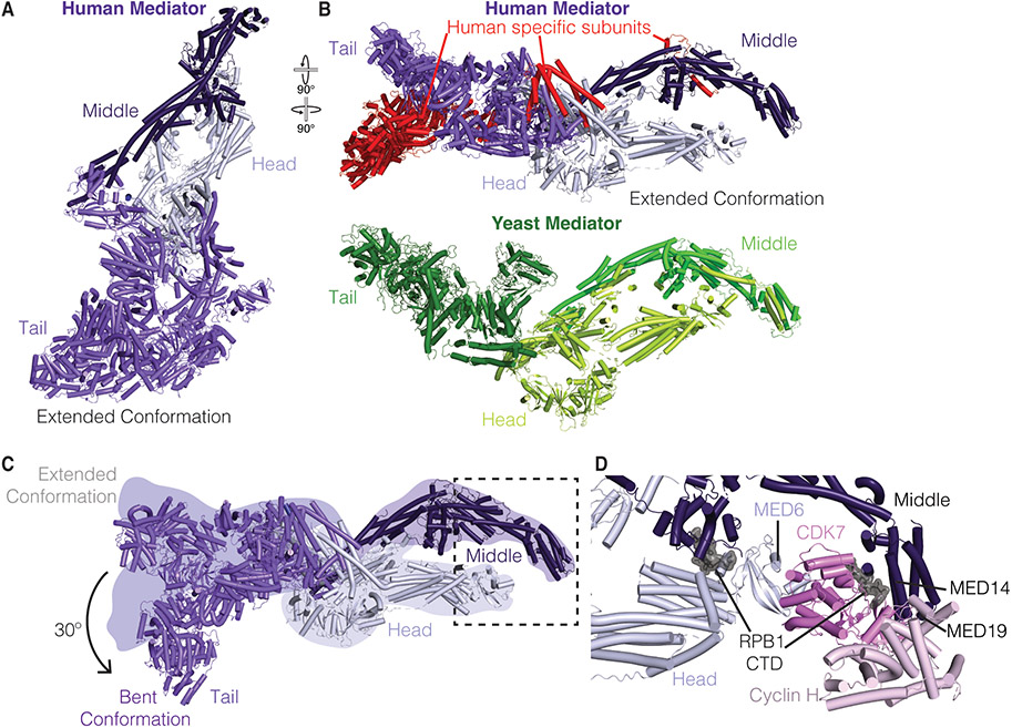
Figure 2. Structural of overview of Mediator and its interactions with TFIIH.
A. Human Mediator in the extended conformation (PDB ID 7EMF)[4]. Head, Middle, and Tail module colored in lilac, dark purple, and violet, respectively.
B. Top: Human Mediator in the extended conformation (PDB ID 7EMF)[4]. Model rotated relative to A. Human specific subunits are colored red. Bottom: Composite structure of yeast Mediator tail (Chaetomium thermophilum, PDB ID 7JMN, Med1, Med24, and unknown subunit Chaetomium thermophilum, PDB ID 6XP5, forest green), and Mediator middle (green) and head (limon) (S. cerevisiae, PDB ID 5OQM)[6,27].
C. Dynamics of the Mediator tail module in extended and bent conformations. Position of extended conformation shown as colored silhouettes. Bent conformation shown in cartoon (PDB ID 7ENJ) [4]. Dotted box indicates binding position of the CAK on Mediator.
D. Interactions of Mediator with TFIIH CAK module (pink). Figure corresponds to boxed region in Panel B. A portion of the RPB1 CTD (grey, surface) was observed in the structure (PDB ID 7LBM)[5].

