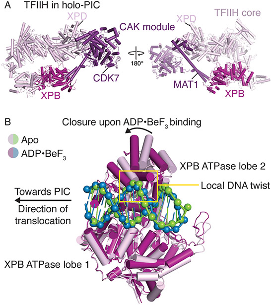
Figure 3. TFIIH architecture and conformational changes in TFIIH XPB.
A. Structural overview of human TFIIH. XPB and the CAK module are colored in fuchsia and purple, respectively (PDB ID 7ENC)[4]. The positions of CDK7, MAT1, and XPD are indicated.
B. DNA translocation by XPB facilitates promoter opening. XPB ATPase lobe 1 in the apo (PDB ID 7NVW, pink and green)[17] and ADP·BeF3 bound states (PDB ID 7NVV, fuchsia and blue)[17] are aligned. Binding of ADP·BeF3 induces closure of the XPB ATPase and propagation of the local DNA twist upstream.

