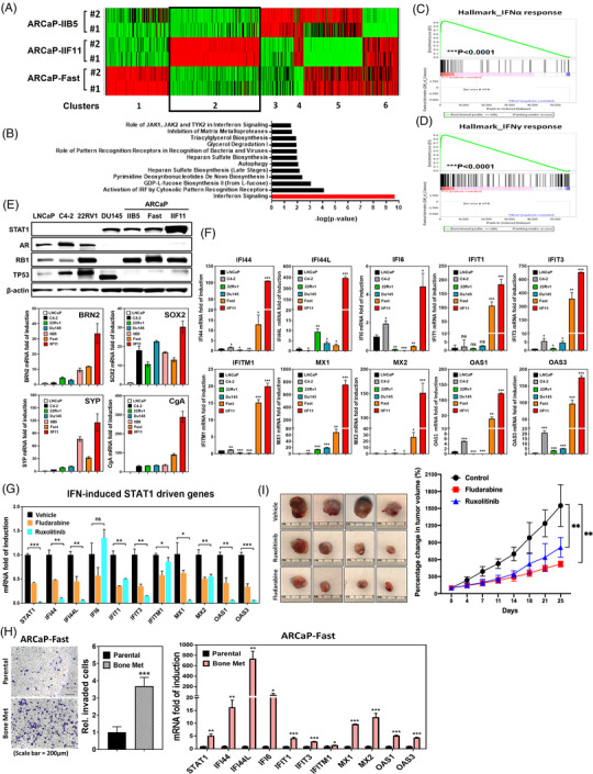
FIGURE 2.

STAT1‐IFIT5 signalling activation emerges during the advanced progression of PCa toward CRPC and NEPC. (A) Heat map illustrating the gene expression profile among three ARCaP sublines‐IIB5, IIF11 and Fast. (B) The ingenuity pathway analysis of significant enrichment of IFN signalling pathway genes in ARCaP‐IIF11, compared to ARCaP‐Fast and ‐IIB5 sublines. (C,D) GSEA identifying enrichment of genes associated with IFNα and IFNγ response in IIF11 subline, compared to Fast and IIB5 sublines. (E) Upper: expression of STAT1, AR, RB1 and TP53 protein levels among screened PCa lines including ARCaP sublines. Lower: screening of BRN2, SOX2, SYP and CgA mRNA levels among PCa lines including ARCaP sublines. (F) Expression level of 10 IFN‐inducible STAT1‐driven genes among PCa lines including ARCaP sublines. (G) Expression of ten IFN‐inducible STAT1‐driven genes in ARCaP‐IIF11 subline treated with fludarabine (1 μM) or ruxolitinib (2.5 μM) for 48 h. (H) Left: expression of ten IFN‐inducible STAT1‐driven genes in ARCaP‐Fast bone metastatic subline, compared to parental line. Right: in vitro invasion assay examining the invasiveness of ARCaP‐Fast bone metastatic line. (I) The impact of fludarabine (20 mg/kg) or ruxolitinib (20 mg/kg) on the sub‐cutaneous ARCaP‐IIF11 tumour growth, compared to vehicle control. (ns = no significant differences, *p < .05, **p < .001, ***p < .0001)
