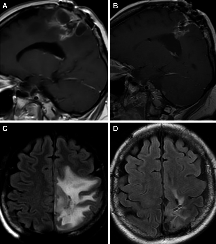
Figure 2. MRIs for patient 10.
Patient 10 had continued disease progression after a second surgical resection as evidenced by increasingly nodular enhancement (A, sagittal magnetic resonance imaging [MRI]) and progressive cerebral edema (B, axial MRI). The patient subsequently initiated ketogenic metabolic therapy, and imaging obtained at the next follow-up evaluation (four months later) showed significant improvement in lesional enhancement (C, sagittal MRI) and markedly reduced cerebral edema (D, axial MRI). Used with permission from Barrow Neurological Institute, Phoenix, Arizona.

