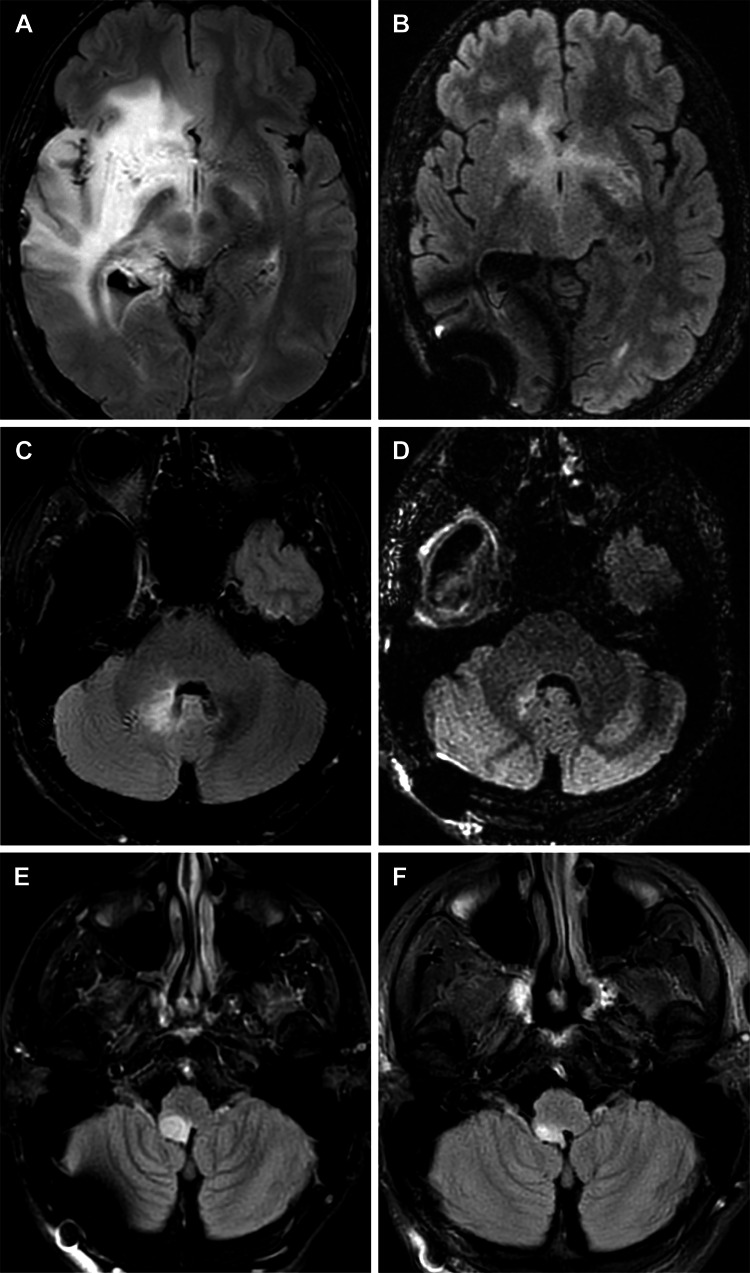
Figure 3. MRIs for patient 1.
Axial magnetic resonance images (MRIs) for patient 1, who underwent repeat surgical resection, show that afterward the patient had continued progression of the tumor within the right insula, thalamus, basal ganglia, basal forebrain (A), and cerebellomedullary region (C). After initiation of ketogenic metabolic therapy, tumor-treating fields therapy, and temozolomide therapy, imaging shows near-complete resolution of the diffuse multifocal fluid-attenuated inversion recovery anomaly in the right insula, thalamus, basal ganglia, basal forebrain (B), and cerebellomedullary region (D). After a period of time during which the patient did not achieve ketosis, subsequent imaging showed progression of the tumor within the right medial temporal lobe and right medulla (E). Metabolic genomic profiling identified the cause of the patient’s inability to achieve ketosis, and he reinitiated ketogenic metabolic therapy. Because of its severity, the cerebellomedullary lesion was treated with Zap-X stereotactic radiosurgery. Follow-up imaging showed that the size of the lesion was greatly reduced (F). Used with permission from Barrow Neurological Institute, Phoenix, Arizona.

