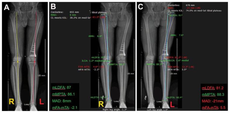
Figure 1.
Assessment and interpretation of extremity alignment studies in a 21-year-old male. A: Extremity alignment revealing a right lower extremity (R) neutral mechanical axis. Observe that the yellow line (Mikulicz line) connecting the center of the hip and the center of the ankle, bisects the knee at its center (yellow asterisk). The left lower extremity (L) has a shift of the mechanical axis to the lateral compartment of the joint. Observe that the Mikulicz line (red) is bisecting the joint (asterisk) on the lateral compartment, which determines a valgus alignment of this lower extremity. B: A software is utilized to determine the joint orientation angles on each one of the lower extremities. Observe that all joint orientation angles around the right knee are marked in green, indicating that they are within normal values. In the left lower extremity, there is a valgus mechanical femoral tibial angle of 5.5 degrees. In this case the deformity is generated at the level of distal femur, as the mechanical lateral distal femur is 81.2 degrees (normal range 85 to 90 degrees). Preoperative planning using digital software provides precise quantification and location of the deformity in the lower extremity.

