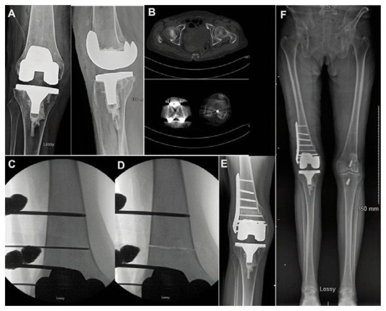
Figure 7.
Illustrative case of torsional deformity. A: Radiographs of a 63-year-old female who underwent a right total knee replacement elsewhere. She had previous valgus alignment and a history of patellectomy many years ago. After her knee replacement she developed significant instability of her extensor mechanism, with recurrent dislocations. She received the indication to revise her total knee replacement. B: In our clinic a computed tomography was performed, and we noticed that she had 24 degrees of internal torsion of the distal femur. She was two months out of her total knee replacement. Instead of revising the components of the knee replacement we proposed an osteotomy to correct the torsional deformity. C: Intraoperative pictures illustrating the orientation of the osteotomy, perpendicular to the axis of the femur; D: The osteotomy has been completed. The femur does not displace because it has been provisionally fixed with an external fixator. E. Final radiograph one year after surgery with complete healing of the osteotomy. Patient has no more symptoms of extensor mechanism dislocation; F: Final alignment study depicting that the osteotomy did not introduce deformity in the coronal plane.

