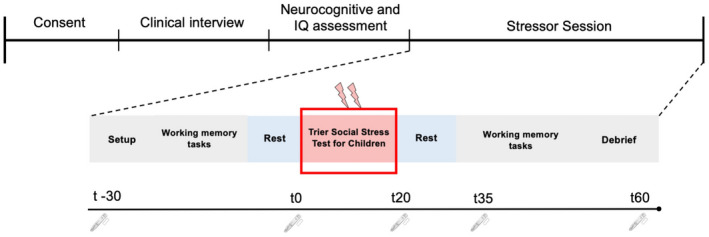
FIGURE 1.

Overall experimental set‐up and design for the stressor protocol. This depicts the order of consent, clinical interviews, and neurocognitive assessment as part of the larger overall study. The stressor session consisted of EEG set up with questionnaires, tasks prior to the stressor (TSST), and a repeat of the same tasks following the stressor. The line underneath corresponds to the time saliva samples were taken for alpha amylase and cortisol. The “t” represents minutes relative to TSST onset. Alpha amylase (fast SNS response) was not assessed at t60, but cortisol (slow HPA response) was
