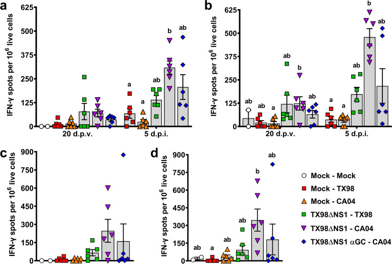
Fig 4.
Cellular responses to TX98 and CA04 measured by IFN-γ ELISPOT assays. IFN-γ production by PBMC collected at 20 d.p.v. and 5 d.p.i. after incubation with UV-inactivated TX98 (a) or CA04 (b) virus particles. IFN-γ production by lung leukocytes isolated at 5 d.p.i. after incubation with UV-inactivated TX98 (c) or CA04 (d) virus particles. Results represent mean IFN-γ spots per 1x10⁶ live cells after subtracting spots counted in unstimulated wells. Differences between treatments were analyzed by Dunn’s multiple comparisons test. A statistically significant difference between two groups is indicated by different letters. Data are represented as mean ± SEM. Symbols represent individual pigs

