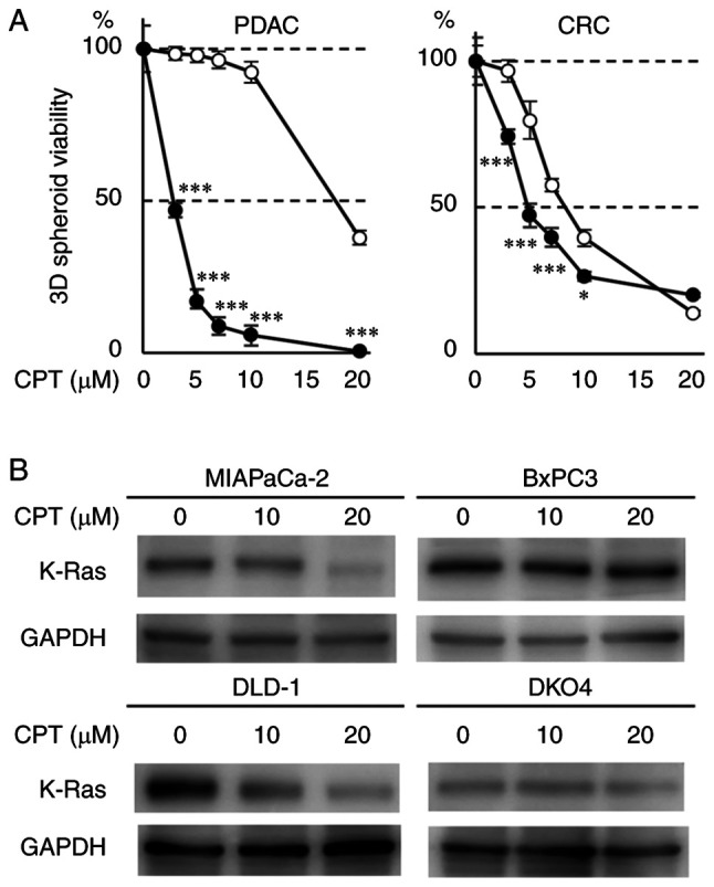
Figure 1.

Effects of CPT on human PDAC (MIAPaCa-2 and BxPC3) and CRC (DLD-1 and DKO4) cell lines. (A) Spheroid viability of PDAC (left) and CRC (right) cell lines treated with CPT. A total of 1×10³ cells were seeded into a 96-well V-bottom plate and incubated with CPT for 72 h in triplicate. Spheroid viability was determined by measuring the ATP content using the CellTiter-Glo 3D assay. Closed and open circles represent the cell lines with mutant KRAS (MIAPaCa-2 and DLD-1) and wild-type KRAS (BxPC3 and DKO4), respectively. Error bars represent standard deviation. *P<0.05 and ***P<0.001 vs. the wild-type KRAS cells treated with the same CPT concentration. (B) Immunoblot analyses of K-Ras protein in cells treated with CPT. A total of 5×10⁵ cells were seeded into poly-HEMA-coated 60 mm dishes and incubated with CPT at 10 and 20 µM for 48 h. GAPDH was used as the internal control. CPT, cryptotanshinone; PDAC, pancreatic ductal adenocarcinoma; CRC, colorectal cancer; KRAS, Kirsten rat sarcoma viral oncogene homolog.
