FIGURE 2.

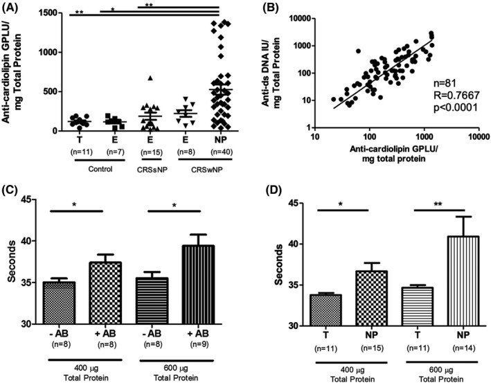
Anti‐cardiolipin in NP and aPTT in vitro Testing. (A) Comparison of anti‐cardiolipin IgG levels in sinonasal tissue from CRS and control patients. Units are shown in immunoglobulin G phospholipid‐binding units (GPLU) normalized to total sample protein. There were significantly higher anti‐cardiolipin IgG in CRSwNP polyp (NP) compared to CRSsNP ethmoid (E), control ethmoid (E), and control turbinate (T) tissue (* p < .05, ** p < .01). (B) Correlation between normalized anti‐cardiolipin IgG antibodies and normalized anti‐dsDNA IgG antibodies. (C) Modified activated partial thromboplastin times (aPTT) using antibodies isolated from control human plasma (‐AB) and lupus‐positive control plasma (+AB). There was a significant increase in aPTT at both 400 µg/mL and 600 µg/mL antibodies derived from +AB samples. (D) Comparison of aPTT times between antibodies isolated from control turbinate (T) and NP tissue. There was a significant prolongation of the modified aPTT by antibodies derived from polyp tissue at both concentrations (* p < .05, ** p < .01)
