Figure 1.

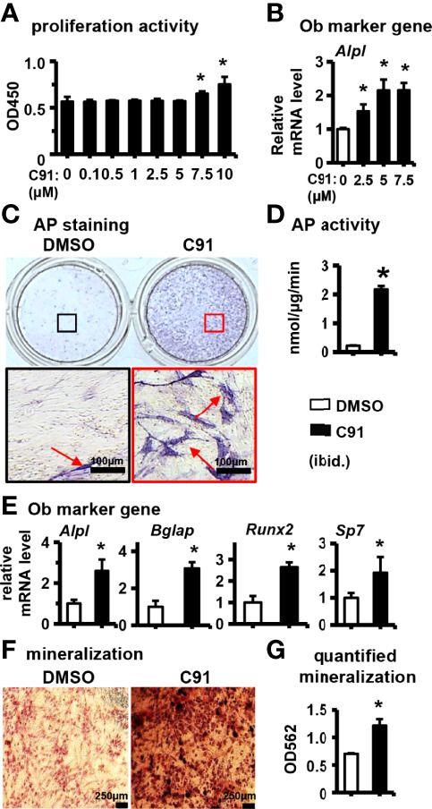
The effect of C91 on ST2 cell differentiation and mineralization. (A) The effect of C91 on the proliferation activity of ST2 cells was detected by CCK8 assay. Results are expressed as relative OD450 values, calculated by normalizing OD450 readings to the mean value of the control group. C91 at concentrations of 0.1–5 μM had no significant effect on the viability of ST2 cells. (B) qRT-PCR detection of mRNA expression of early osteogenesis marker gene Alpl. ST2 cells were treated with C91 (5 μM) for 3 days, followed by AP staining (C) analysis and AP biochemical quantification (D) analysis. Red arrows indicate alkaline phosphatase staining positive cells. Scale bar = 100 μm. (E) The mRNA expression levels of osteogenic target genes were detected by qRT-PCR. (F) ST2 cells were treated with C91 for 14 days, and the osteogenic induction medium was changed every two days in between. Alizarin Red S staining was performed (original magnification, x4). Red arrows indicate mineralized nodules, scale bar = 250 μm. (G) is the quantitative analysis result of panel (D) Results are presented as mean ± SD (n = 3 per group). * indicates P < 0.05, compared with the DMSO group.
